![[ICON: To the NSR page]](../../graphics/nsrlogo.gif)
![[ICON: To the NSR page]](../../graphics/nsrlogo.gif)
![[ICON: To the RIM page]](../../graphics/rimlogo.gif)
X-ray powder diffraction is a powerful technique for the identification of solid state samples. The underlying crystal structure of matter will, in principle, give rise to a unique X-ray powder diffraction pattern (which expresses the existence of lattice planes in materials) for each pure compound and for each individual component in a mixture of compounds.
Powders generally contain a very large number of randomly oriented microcrystals, each of which diffracts independently, yielding continuous cones of Bragg reflections that are recorded as lines on an X-ray powder diffractometer when these cones are crossed by the scanning counting equipment. Each line corresponds to a specific diffraction angle at which the X-ray beam is reflected by a specific set of lattice planes in the powder. Because of the random orientation of the microcrystals in the powder, all possible lattice planes will reflect sooner or later.
The combination of the unique set of angles (q) at which a powder reflects X-rays and the corresponding intensities, yields a fingerprint that is unique for the crystal structure (and hence its polymorph) of each single component. The positions of the peaks in an X-ray powder diffraction pattern are directly related to the dimensions of the unit cell, which is defined by six so called cell parameters. The intensities corresponding to the peaks are related to the contents of the unit cell (the atomic positions, or better: the electron density distribution).
Different polymorphs have different crystal structures due to a different packing of the molecules in the lattice. This results in a different crystal symmetry and/or unit cell parameters which directly influence the reflection characteristics of crystals or powders. A different polymorph will in general diffract at a different set of angles and will give other values for the intensities. Therefore X-ray powder diffraction can be used to identify different polymorphs or a mixture of polymorphs in a reproducible and reliable way.
Experimental setup requires that, if a reflection is diffracted when the incoming beam forms an angle q with a certain lattice plane, the reflected beam is recorded at an angle 2q.
Although 2q values are part of the printed output, normally peaks are characterized by their unique 'DI-values': D stands for d-value (the distance between lattice planes), and I for intensity.
The d-values are preferred because they are independent of the type of radiation used (wavelength independent), in contrast with the 2q values. They are calculated using Bragg's Law: 2 d sinq = nl.
Because of differences in experimental conditions, normally relative intensities are used: the strongest peak is set to 100, all other intensities are scaled accordingly.
When interpreting X-ray powder diffraction patterns, several effects should be taken into account. In order of importance: peak intensities can be influenced considerably by 'preferred orientation' (i.e. the particles are not distributed randomly) and a variety of other effects (with lesser influence); their widths are rather dependent on particle size (overly small particles will result in line broadening); and, as a minor effect, their positions can be slightly influenced by the sample thickness (i.e. changes in height adjustment), sample roughness (i.e. smoothness of the sample surface) and sample 'transparancy' (i.e. how deep the radiation will penetrate: an absorption-related effect). Only some of these effects can be minimized by careful sample preparation.
The X-ray powder diffraction pattern of a mixture of two or more components is, to a large extent, the weighted sum of the individual patterns. However, differences in particle sizes and absorption effects can have substantial influence. This means that some of the effects mentioned are (or can be) 'anisotropic', depending on how large these differences are.
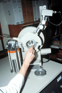
 Philips PW1820 Diffractometer
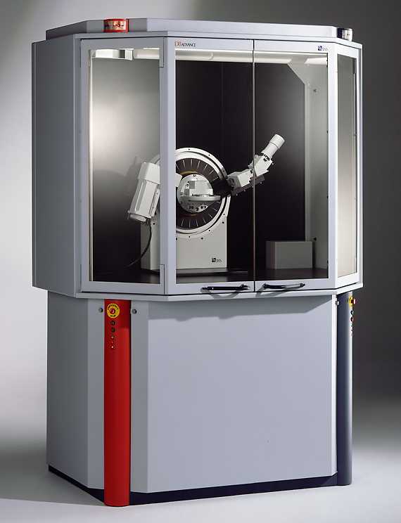
 Bruker D8 Advance