1) SCANNING THE POWDER XRD FILM
A flat bed scanner with a minimum resolution of 300 DPI and some sort of transparency adapter is needed. Many scanners come equipped with transparency adapters, but even an inexpensive scanner can be converted with a transparency adapter made out of a high frequency fluorescent light
Tips for obtaining a good scan:
a) make certain that the diffraction pattern axis is parallel to the (long) axis of the scanner. While an image can be rotated in most image processing programs, this will introduce distortion into the image
b) Set the initial image defaults to the minimum brightness and gamma that will show the diffraction lines. Low contrast should be used.
c) Save the image in *.TIF or a similar uncompressed format
d) While the programs used in this tutorial will work with color images, it is best to set the scan to monochrome 256 shades of gray
Here are some examples of the raw images after scanning:
..................................................................................................................................................................................................................................................
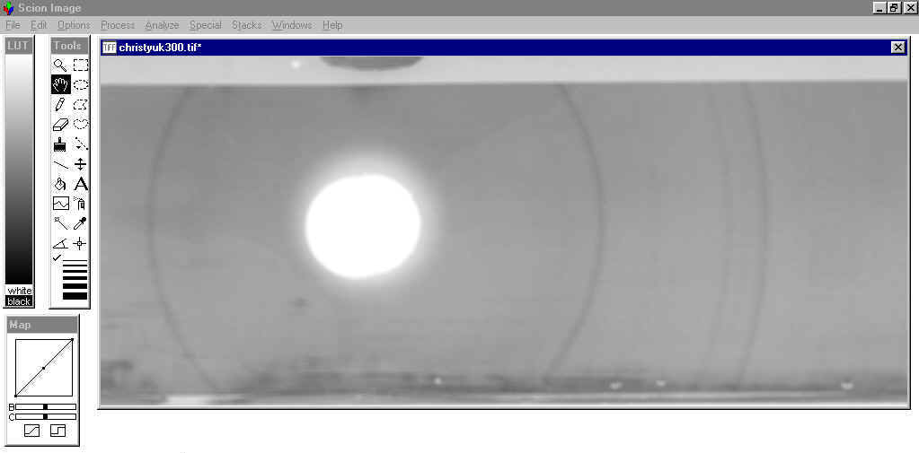
This image is an unknown vanadium-iron oxide from the Christy Vanadium Mine in Arkansas. The diffraction pattern was prepared using Cu X-rays and had an extremely high background from the Fe fluorescence. The film was so dark that the inner emulsion was removed to lighten the image. After adjusting the gamma and brightness, the diffraction lines were clearly visible. This image, representing somewhat of a "worst-case scenario" will be used in the rest of the tutorial.
The image below was prepared with a high frequency fluorescent light as a transparency adapter. The film was scanned by placing it in a plastic film preserver sleeve attached to a black piece of foamboard. The fluorescent light was placed on top of an opening in the foamboard.
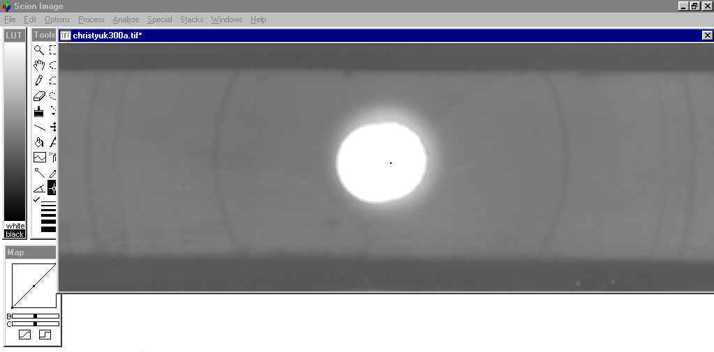
The image below was taken using a film scanning device that came with the same scanner. There are subtle differences in the background that actually make this a better scan despite being visually inferior to the scan above.
......................................................................................................................................................................................................................................................
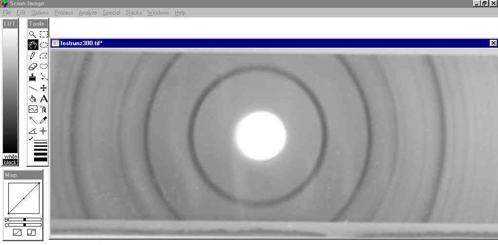
This image is the mineral ferrostrunzite from Clear Springs Mine Bartow, Florida. The diffraction pattern was prepared using a Gandolphi camera and Co X-rays. The image has a low background and was scanned with minimal adjustment of the gamma and brightness. A large sphere mount (0.7mm) was used and this resulted in a very dark, but broad diffraction line on the film.
.....................................................................................................................................................................................................................................................
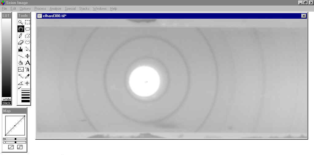
This image is an undescribed mineral species related to ferrostrunzite from CF Hardee Mine in Florida. The film was also prepared using Co X-rays and a Gandolphi camera. The sample was mounted as a sphere <0.2mm in diameter.